Pages

Search This Blog

Chitika

Showing posts with label ORTHOPAEDICS. Show all posts

Chopart's fracture


The foot is generally divided into the
    • Hindfoot
      • Calcaneus
      • Talus
    • Midfoot
      • Cuboid
      • Navicular
      • Three cuneiforms
    • Forefoot
      • Metatarsals
      • Phalanges
    • The articulation between the hindfoot and the midfoot (midtarsal joint) is frequently referred to as Chopart’s joint
      • Named after surgeon who performed amputations at the calcaneocuboid, talonavicular joint
    • The articulation between the midfoot and the forefoot is referred to as the Lisfranc joint.
      • Named after French surgeon Francois Chopart (1743–1795) who performed amputations of the foot at this level
      • This type of amputation renders the ankle joint unstable as almost all of the points of insertion of the ankle tendons have been remove
  • All dislocations of the foot are relatively uncommon with the Lisfranc fracture-dislocation being the most common
  • Most are due to falls from a height or motor vehicle accidents
  • Males are more likely to have foot dislocations than females
  • Chopart Fracture
    • Chopart fracture-dislocation involves the midtarsal joints (talonavicular and calcaneocuboid joints)
    • Typically caused by falls from a height, motor vehicle accidents and severe twisting injuries such as can occur in basketball players who land on a plantar-flexed and inverted foot
    • Usually result from severe trauma
    • Most commonly, there is medial displacement of the distal fragments (80%)
    • The foot is displaced inward and upward
      • But displacement in other directions can occur
      • Eversion injuries result in lateral dislocations
    • There are frequently associated fractures of the calcaneus, cuboid and navicular
    • A small percentage are open
    • The talus remains in the ankle mortise


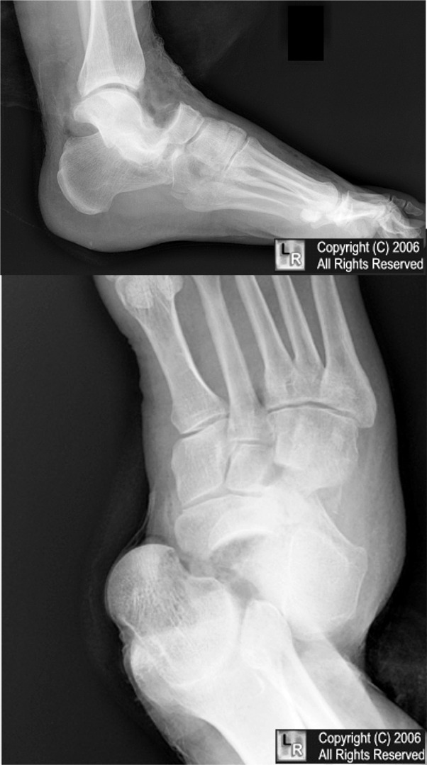
Chopart's fracture dislocation.
Black arrow points to talus which is dislocated from navicular (yellow
arrow) at talonavicular joint. Calcaneus (blue arrow) is dislocated from the cuboid (red arrow), which is also fractured. The dislocation is at the calcaneocuboid joint. This is an uncommon dislocation.
The forefoot is usually displaced medially rather than laterally as in this case.
  • Prognosis
    • Prompt reduction and early range of motion generally result in favorable outcome
    • High impact injuries with greater soft tissue compromise and associated fractures worsen prognosis
.

Diagram of Chopart's fracture-dislocation from Radiographics.

Carpal tunnel syndrome


This condition was previously most commonly found
in women, 30 to 60 years of age. With the
increasing use of computers, Carpal Tunnel syndrome
is now seen with increasED frequency in all ages,

Phalen's test


Place the backs of both of your hands together and hold the wrists in forced flexion for a full minute. (Stop at once if sharp pain occurs) . If this produces numbness or "pins and needles" along the thumb side half of the hand, you most likely have Median nerve entrapment (Carpal Tunnel Syndrome). Examination by a health care professional familiar with these conditions is the way to be sure of the diagnosis and get proper treatment.

Hammer toes


Hammer toes come in all shapes and sizes. Hammer toes can be found to affect one of the toes or all of the toes simultaneously. The name, hammer toe comes from the way the toe hits or hammers on the floor with each step. The primary deformity seen in a hammer toe is found at the PIPJ (proximal interphalangeal joint) which is the first or more proximal of the two joints of the toe. A

Bohler's angle ( Tuber joint angle )


-Bohler's angle also called as the Tuber joint angle, measures the angular relationship between talus and calcaneum. This angle is formed by two lines . first line is drawn from the posteriosuperior margin of the calcaneal tuberosity through the tip of the posterior facet of the subtalar joint. second line is drawn from the tip of the posterior facet through the superior margin of the anterior process of the calcaneum.
-Normally this angle ranges between 20 and 40 degrees.
-Flattening of this angle is a classic x-ray sign of depressed fracture of calcaneum. 
P.S : The image is not my original work and has been taken from the wikimedia commons project and the author is Gilo1969.

Gallows traction


*This is used in infants and children with femoral fractures.

*Indications Gallows Traction
- Child must weigh less than 12 kg
- Femoral fractures
- Skin must be intact

*Both the fractured and the well femur are placed in skin traction and the infant is suspended by these from a special frame. Vascular compromise is the biggest danger. Check the circulation twice daily. The buttocks should be just off the bed.

Knuckle bender splint


*These are used in ULNAR NERVE PALSY.

Aeroplane splint


*The above picture shows the AEROPLANE SPLINT, which is used in brachial plexus injury.

Hip spica


*A hip spica is used in femoral fractures.

Patellar Tendon bearing brace


*A patellar tendon bearing brace is used in Tibial fractures.

Ilizarov External Fixator


The Ilizarov apparatus is named after the orthopedic surgeon, Gavril Abramovich Ilizarov, from Siberia who pioneered the technique. It is used in surgical procedures to lengthen or reshape limb bones; treat complex and/or open bone fractures; and in cases of infected non-unions of bones that are not amenable with other techniques.

Professor Gavril Abramovich Ilizarov invented this procedure in the 1950s after having to treat orthopedic conditions in the Kurgan region of Siberia. The procedure, and the first apparatus he designed for it, was inspired by a shaft bow harness on a horse carriage.[1] Originally bicycle parts were used for the frame.

This novel technique was introduced to the West in the 1980s, predominantly via Italian surgeons. It gained popularity in the 1990s, and has been used successfully by many surgeons throughout the world. In most developing countries it is a highly specialised technique used mainly for deformity correction by experienced surgeons due to its complexity. Further development of the ring construct led to the Taylor Spatial Frame which is more versatile and far easier to use, but very costly. Though nowadays intramedullary limb lengthening devices are also available, they are not suitable for deformity correction of bones.

The device is a specialized form of external fixator, a circular fixator, modular in construction. Stainless steel rings are fixed to the bone via stainless heavy-gauge wire (called "pins" or Kirschner wires). The rings are connected to each other with threaded rods attached through adjustable nuts. The circular construction and tensioned wires of the Ilizarov apparatus provide far more structural support than the traditional monolateral fixator system. This allows early weightbearing. The frame can be used to support a fractured limb, but it is most commonly used to correct deformity through distraction osteogenesis.

The procedure consists of an initial surgery, during which the bone is surgically fractured and the ring apparatus is attached. As the patient recovers, the fractured bone begins to grow together. While the bone is growing, the frame is adjusted by means of turning the nuts, thus increasing the space between two rings. As the rings are connected to opposite sides of the fracture, this adjustment, done four times a day, moves the now-healing fracture apart by approximately one millimeter per day. The incremental daily increases result in a considerable lengthening of the limb over time. Once the lengthening phase is complete, the apparatus stays on the limb for a consolidation period. The patient is able to fully weight bear on the Ilizarov frame, using crutches initially and pain is lessened. Once healing is complete, a second surgery is necessary to remove the ring apparatus. The result is a limb that is significantly longer. Additional surgery may be necessary, in the case of leg lengthening, to lengthen the Achilles tendon to accommodate the longer bone length. The major advantage of this procedure is that because the apparatus provides complete support while the bone is recovering the patient can remain active aiding recovery.

A further use is of bone transport, whereby a defect in a long bone can be treated by transporting a segment of bone, whilst simultaneously lengthening regenerate to reduce the defect and finally dock with the other segment, producing a single bony unit.

While the Ilizarov apparatus is minimally invasive (no large incisions are made,) it is not free of complications. Pain is common and can be severe, but is treatable with analgesics. Careful attention to cleaning and hygiene is necessary to prevent pin site infection. Other complications include swelling and muscle transfixion.

The Ilizarov method is widely used to treat complex and/or open bone fractures. This method is preferred over conventional treatment options (such as internal fixator or cast) where there is a high risk of infection or the fracture is of such severity that internal fixators are unworkable.

Haglund's deformity (Pump Bump)


*A Haglund deformity, or pump bump, is caused by chronic inflammation of the adventitious superficial pretendinous Achilles bursa that separates the Achilles tendon from the overlying skin.

*According to Jones, this bursa is present in about 50% of patients. This pretendinous bursitis usually is caused by chronic irritation from a shoe heel counter, and modification of shoe wear usually relieves symptoms. This deformity usually occurs in young women in their 20s or 30s. Surgery is infrequently required.

*The following technique is recommended if conservative measures fail :

1. Place the patient prone. After administration of general or local anesthesia, make a longitudinal lateral incision 1 cm lateral to the Achilles tendon, extending distally from 3 to 4 cm proximal to the superior tuberosity of the calcaneus to 2 to 3 cm distal to the superior tuberosity of the calcaneus.

2. Plantar flex the ankle joint and by sharp and blunt dissection, identify the Achilles tendon.

3. Place a right-angle retractor between the Achilles tendon and posterior and superior borders of the calcaneal tuberosity. With the foot plantar flexed, this usually affords enough exposure to remove the superior border of the calcaneal tuberosity without raising any of the Achilles tendon off the calcaneus. However, the Achilles tendon has such an extensive insertion into the posterior and plantar aspect of the calcaneal tuberosity that raising a 1- to 2-cm-long portion of the tendon may be necessary to resect the bone adequately.

4. Remove the superior aspect of the tuberosity with a microsagittal saw or an osteotome. Placement of several drill holes along the proposed osteotomy site makes this resection easier.

5. If an area of ossification remains, split the Achilles tendon in a coronal plane distally with the anterior third or half to free up enough to excise the calcified tendon.

6. Lavage the wound and close in layers.

7. Apply a well-padded, short leg, non–weight bearing cast with the ankle in approximately 20 degrees of plantar flexion.

AFTER TREATMENT:
*The cast and sutures are removed at 3 weeks. The sutures may be removed earlier if indicated, but the non–weight bearing cast remains on for 3 weeks. Then a removable weight bearing cast boot is applied, and active plantar flexion and dorsiflexion exercises are begun. It is important in the preoperative counseling to explain to a young woman with a pump bump that it might be 3 to 6 months before she can wear a stylish shoe and that there is no guarantee that she will ever be able to do so comfortably.

Pathophysiology of Paget's disease (Osteitis deformans)


BASIC PHYSIOLOGY OF NORMAL BONE RESORPTION AND FORMATION :


*Osteoclast maturation is regulated by various factors, such as Receptor Activator of Nuclear Factor-kappaB (NF-kappaB) Ligand (RANKL). RANKL can exist as a soluble form and binds to the osteoclast receptor RANK.
*This binding activates osteoclast differentiation via the translocation of NF-kappaB into the nucleus by intermediates such as Tumor Necrosis Factor (TNF) Receptor-Associated Factor 6 (TRAF6).
*RANKL signals can be blocked by a soluble RANKL-binding decoy receptor, osteoprotegerin (OPG), produced by osteoblasts.
*Vacuolar H+-ATPase pump (H+), matrix metalloproteinase (MMP), Cathepsin K and Tartrate-Resistant Acid Phosphatase (TRAcP) are involved in osteoclast activity.

*This picture above helps us in understanding the pathophysiology behind PAGET'S disease :

*PATHOPHYSIOLOGY OF PAGET'S DISEASE:
- The principal abnormality in Paget disease is the increased number and activity of osteoclasts. 
- Pagetic osteoclasts are large, increased 10- to 100-fold in number, and have a greater number of nuclei (as many as 100 compared to 3–5 nuclei in the normal osteoclast). 
- The overactive osteoclasts may create a sevenfold increase in resorptive surfaces and an erosion rate of 9 microg/d (normal is 1 microg/d).
- Several causes for the increased number and activity of pagetic osteoclasts have been identified: 
(1) Osteoclastic precursors are hypersensitive to 1,25(OH)2D3
(2) Osteoclasts are hyperresponsive to RANK ligand (RANKL), the osteoclast stimulatory factor that mediates the effects of most osteotropic factors on osteoclast formation; 
(3) Marrow stromal cells from pagetic lesions have increased RANKL expression; 
(4) Osteoclast precursor recruitment is increased by interleukin (IL) 6, which is increased in the blood of patients with active Paget disease and is overexpressed in pagetic osteoclasts; 
(5) Expression of the proto-oncogene c-fos, which increases osteoclastic activity, is increased; and 
(6) The antiapoptotic oncogene Bcl-2 in pagetic bone is overexpressed. Numerous osteoblasts are recruited to active resorption sites and produce large amounts of new bone matrix. As a result, bone turnover is high and bone mass is normal or increased, not reduced.

- The characteristic feature of Paget disease is increased bone resorption accompanied by accelerated bone formation. 
- An initial osteolytic phase involves prominent bone resorption and marked hypervascularization. 
- Radiographically, this manifests as an advancing lytic wedge, or "blade of grass" lesion. 
- The second phase is a period of very active bone formation and resorption that replaces normal lamellar bone with haphazard (woven) bone. 
- The mosaic pattern of woven bone is structurally inferior and can bow and fracture more readily. 
- At the same time, fibrous connective tissue may replace normal bone marrow. 
- In the final sclerotic phase, bone resorption declines progressively and leads to a hard, dense, less vascular pagetic or mosaic bone, which represents the so-called burned-out phase of Paget disease. 
- All three phases may be present at the same time at different skeletal sites.

*Acutely marginated bone demineralization during lytic phase in skull - OSTEOPOROSIS CIRCUMSCRIPTA.
*Acutely marginated demineralization of long bones - BLADE OF GRASS SIGN and FLAME SHAPED MARGIN.
*Mixed lytic and sclerotic phase in spine - PICTURE FRAME VERTEBRAE
*Mixed lytic and sclerotic phase in skull - COTTON WOOL SKULL.

Bumper fracture


*A bumper fracture is a compression fracture of the lateral tibial condyle due to a forceful valgus stress applied to the knee.

*The name is derived from the fact that a car bumper hitting the lateral aspect of the knee when the leg is firmly planted on the ground is one of the most common causes of this type of injury.

*If the medial collateral ligament remains intact, the lateral femoral condyle is forced down on the lateral tibial condyle and this causes a compression fracture.

*Older patients with osteoporosis are the most prone to this type of injury. There may be a hemarthrosis, and the lateral tibial plateau will be tender.

*Compression fracture of the lateral tibial plateua, resulting in separation at the margin of the plateau or depression of the central portion of the artucular surface.

*A valgus injury of the knee may result in a fracture of the lateral tibial plateau. At times these fractures are difficult to identify and are obvious only on oblique radiographs.

*The fractures consist of either a vertical split through or a depression of a portion of the joint surface. There may be an associated fracture of the neck of the fibula.

*When the fracture involves the lateral margin of the plateau or is associated with a fracture of the fibula, an accompanying disruption of the medial collateral ligament is likely.

*Treatment: Stabilisation and fixation where required.


Pins, screws and Prosthesis used in Fracture neck of Femur


*When a child presents with fracture neck of femur which is less than 3 weeks old, then the fracture is managed by CLOSED REDUCTION and INTERNAL FIXATION with Austin Moore pins and Knowle's pins.

*Austin Moore pin and Knowle's pin shown in the picture above.


*X-ray of a case of Fracture neck of femur, fitted with Austin Moore pins.


*Illustration showing the Knowle's pins used in the management of fracture neck of femur.


*X-ray of a case of fracture neck of femur managed by knowle's pins.


*When an adult whose age is less than 60 years presents with a fracture neck of femur, less than 3 weeks old, then the fracture is managed by Closed/open reduction and internal fixation by MULTIPLE CANNULATED CANCELLOUS SCREWS (shown in the picture above)


*When an adult whose age is more than 60 years presents with fracture of neck of femur, less than 3 weeks old, then the next step is to assess his hip and take a decision. If there is preexisting arthritis in the patient, then Total Hip Replacement is the treatment of choice.
- But if the hip is otherwise normal, then Hemireplacement arthroplasty is done. A.M.Prosthesis is one of several prostheses used in Hemireplacement arthroplasty.

Basic Hip fractures

Congenital scoliosis


*Scoliosis is the sideways curvature of the spine.

*It is classified into two major types. They are Non-structural (transient) and Structural (permanent) types of scoliosis

*Non-structural scoliosis is again divided into POSTURAL scoliosis, COMPENSATORY and SCIATIC scoliosis.

*Structural scoliosis is divided into IDIOPATHIC, CONGENITAL and PARALYTIC scoliosis.

*Overall POSTURAL scloliosis is the most common type of scoliosis.

*IDIOPATHIC scoliosis is the most common type of permanent scoliosis.

*CONGENITAL SCOLIOSIS :
- This type is always associated with some form of radiologically demonstrable anomaly of the vertebral bodies. These are

A. HEMIVERTEBRAE - Only One half of the vertebra grows


B. BLOCK VERTEBRAE :- Two vertebral bodies are fused. 


C. UNSEGMENTED BAR : - A bar of bone joining two adjacent vertebrae on one side, thereby preventing growth on that side. 


*Shown below are the Plain radiographs of a pediatric patient with congenital scoliosis. (A) Posterior--anterior and (B) lateral views of the spine, with multiple congenital vertebral anomalies including hemivertebrae at thoracic and lumbar spine and block vertebrae at lumbar spine.

Cobb's angle


*Cobb's angle, a measurement used for evaluation of curves in scoliosis on an AP radiographic projection of the spine (Fig.1).

*When assessing a curve the apical vertebra is first identified; this is the most likely displaced and rotated vertebra with the least tilted end plate. The end/transitional vertebra are then identified through the curve above and below. The end vertebra are the most superior and inferior vertebra which are least displaced and rotated and have the maximally tilted end plate.

*A line is drawn along the superior end plate of the superior end vertebra and a second line drawn along the inferior end plate of the inferior end vertebra.

*If the end plates are indistinct the line may be drawn through the pedicles. The angle between these two lines (or lines drawn perpendicular to them) is measured as the Cobb angle.

*In S-shaped scoliosis where there are two contiguous curves the lower end vertebra of the upper curve will represent the upper end vertebra of the lower curve. Because the Cobb angle reflects curvature only in a single plane and fails to account for vertebral rotation it may not accurately demonstrate the severity of three dimensional spinal deformity.

*As a general rule a Cobb angle of 10 is regarded as a minimum angulation to define scoliosis.

Supports used in Scoliosis


*The above picture shows the MILWAUKEE brace, which is named after the city of Milwaukee where it was designed.


*It is most more acceptable than other braces.


*This is a body cast with a turn-buckle in between. The tightening of the turn-buckle stretches the concave side of the curve, thus correcting the deformity.

Critical angle of Gissane


*In 1947, Gissane described his critical angle or crucial angle.

*He noted a distinct angular cortical platform that parallels the lateral process of the talus on lateral radiographic projection.

*This cortical density represents the dense subchondral bone lying beneath the posterior, anterior and middle facets.

*The angular measurements vary from 130 to 145 degrees, with an average of 130 degrees.

*During an axial impaction load, the lateral process of the talus is driven through the posterior facet in a wedge like manner that facilitates the primary fracture. This extends from the lateral cortical vertex of the crucial angle and exits plantarly through the neutral triangle.

*The initial fracture, as described by Essex Lopresti, is located on the anterior distal lip of the posterior facet and connects to the primary fracture line. This fracture extends through the facet, splits it into one or multiple fragments, and impacts the lateral portions into the body.

*The crucial angle reveals the angular relationship of the calcaneal facets and should appear identical when taken bilaterally.

*Unlike Bohler’s angle, which may be aberrant with displaced extraarticular and intraarticular fractures, the crucial angle is more specific for intraarticular distortion.


 *In the above second figure: Lateral radiograph of the calcaneus shows compression (light blue arrows) and traction (yellow arrows) trabeculae, with the neutral triangle (brown triangle) in between with sparse trabeculae. The thickened cortical or thalamic portion of the bone supporting the articular facets is shown (T). The critical angle of Gissane (G) and the Boehler angle (B) have also been drawn in. The Boehler angle is normally 20°–40°.
Related Posts Plugin for WordPress, Blogger...